Return to flip book view
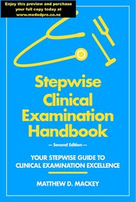
Stepwise Clinical Examination Handbook— Second Edition —YOUR STEPWISE GUIDE TO CLINICAL EXAMINATION EXCELLENCEMATTHEW D. MACKEY
Stepwise Clinical Examination Handbook— Second Edition — Your Stepwise Guide to Clinical Examination Excellence Dr. Matthew D. Mackey MBChB, DCH, PGDipClinEd, FHEA Advanced Trainee RACP and RCPA 2019 New Zealand
!Copyright © 2019 M. D. Mackey First edition published 2018 This edition published December 2019 by MedEdPRO, NEW ZEALAND Cover design by M. D. Mackey This book is copyright. Except for the purpose of fair review, no part of this book may be reproduced or utilised in any form or by any means, electronic or mechanical, including photocopying, recording or by any information storage and retrieval system, without permission in writing from the author, MedEdPRO or its agents. ISBN: 978-0-473-47907-7 DISCLAIMER This book and the content contained within are primarily for educational purposes and do not replace professional medical advice. No responsibility is assumed by the author, MedEdPRO or any associated parties, for any clinical or academic shortcomings, injury or damage to persons or property, from the use of the content or methods contained within this book.
!Peer ReviewersDr. Zaheer BHIKOO MBChB, FRACP Specialist Respiratory Physician, Caboolture Hospital, AUSTRALIA Dr. Jessie MA MBChB, DipOMG Advanced Trainee General Medicine, Waikato Hospital, NEW ZEALAND Dr. Ahmad Nuriman Ahmad NIZAR MBChB Advanced Trainee Cardiology and General Medicine, Waikato Hospital, NEW ZEALAND Dr. Matthew PHILLIPS BScH, MSc, BMBS, FRACP Specialist Neurologist, Waikato Hospital, NEW ZEALAND Dr. Jade TAMATEA MBChB, FRACP, PhD Specialist Endocrinologist, Waikato Hospital, NEW ZEALAND Senior Lecturer, University of Auckland, NEW ZEALAND Dr. Eddie Kuok Chuin TAN BMBS, MRCP, CCT Renal Med, FRACP Specialist Nephrologist, Waikato Hospital, NEW ZEALANDMedEdPRO3
!IntroductionBeing proficient in the clinical examination is one of the traditional skills deemed necessary of a competent physician. However, developing proficiency in the clinical examination is a complex process which requires repetitive practice and quality feedback. Ideally, such repetitive practice and quality feedback is guided by an experienced physician skilled in the arts of clinical examination and education. Unfortunately, given the dynamic and busy nature of the clinical environment, this ideal is not always possible. As such, ‘Stepwise Clinical Examination Handbook’ has been created to be the ‘guide-on-the-side’ for trainee health professionals looking to develop proficiency in the physical examination of an adult patient, in addition to being a quick reference guide for more experienced physicians seeking a refresher in the various clinical examinations. The checklist components of this book have been designed for use during the formative feedback process. It is envisioned peer examiners and clinical educators will complete the checklist as each recommended examination step is performed, in addition to using the checklist to identify aspects of an individual’s examination technique in need of refinement. The relevant checklist should be reviewed within the feedback process as an objective record of the individual’s performance. This book is not designed, nor claims, to be the definitive guide in the physical examination of the adult patient. As such, it is expected once the user gains sufficient experience, confidence and proficiency in the various clinical examinations detailed within this book, alongside considering, and incorporating where appropriate, viewpoints from other sources (e.g. colleagues, mentors, educators and print), the user will feel free to modify their examination approach from that listed to a more individualised style. In addition, based on the author’s experiences, the differential diagnoses of important clinical signs/conditions have been listed for reference (part II), along with ‘difficult-to-master’ examination techniques being described in detail (part III). As will become evident as you start to use the book, a gold diamond symbol signifies the specific examination techniques which are explained in detail in the glossary section.MedEdPRO5
!Cardiovascular examinationRespiratory examinationGastrointestinal examinationRenal (+/- transplant) examinationRheumatological hand examinationRheumatoid arthritis examinationSystemic sclerosis examinationBack and proceed examinationNeck and proceed examinationThyroid biochemical status examinationAcromegaly examinationCushing’s syndrome examinationHypopituitarism examinationEye and proceed examinationCranial nerve examinationUpper limb neurological examinationLower limb neurological examinationAbdomen and proceed examinationGait and proceed examinationExtrapyramidal examinationCerebellar examinationMyotonic dystrophy examinationPart I - Clinical Examinations10121618202224262830323436384042441446CONTENTS485052Speech and Dysarthria examinationsDysphasia examinationHigher functions / centres examination545658MedEdPRO6
!Part II - Quick Reference DifferentialsCardiac murmursMurmur dynamic manoeuvresDigital clubbingRespiratory signsInterstitial lung diseasePleural effusionAbdominal organomegalyNon-palpable apex beatHypopituitarismNeuro-ophthalmologic signsNeuro-ophthalmologic cranial nerve palsiesPeripheral neuropathyCerebellar disorders6262636465656663Glossary77Index108Part III - Glossary of Recommended Examination Techniques6869707273MedEdPRO7
PART IClinical ExaminationsMedEdPRO9
!CARDIOVASCULAR EXAMINATIONGENERAL 1W" - Wash hands, either in examination room or outside - if outside ensure you inform your patient and examiner(s).2I " - Introduce self and role to patient. Ensure patient can hear you adequately. Briefly explain what you plan to do. 3P - Pain - Enquire about presence/location of pain. Advise patient to alert you to any discomfort during the exam.4E - End-of-bed observations - patient (appearance, position, breathing pattern) and environment (medications, equipment).5R - Reposition patient supine on bed with upper body elevated 45° and gender-considerate chest exposure.UPPER LIMBS6Inspect nails, fingers, dorsal and palmar surfaces of hands (clubbing - ! Schamroth’s Sign, colour, tar staining, SBE lesions - splinter haemorrhages/Janeway lesions/Osler’s nodes, arachnodactyly, tendon xanthomata, temperature). 7Inspect upper limbs (vessel harvesting scars, IV track marks, bruising).8Palpate radial pulse for rate and rhythm.9Simultaneously palpate both radial pulses (radio-radio delay of aortic coarctation) - consider testing for radio-femoral delay.10Examine for ! Water Hammer Pulse (differential includes aortic regurgitation).11Perform ! Manual Blood Pressure Measurement - ideally on each arm, then ! Postural Blood Pressure Measurement.FACE12Inspect face, eyes and conjunctiva (malar flush, xanthomata, conjunctival pallor, corneal arcus). 13Perform ! Fundoscopy (Roth’s spots, diabetic & hypertensive changes). **Unlikely to perform in short case/OSCE setting.**14Inspect mouth and oropharynx with a torch (central cyanosis, dentition, arch palate).NECK15Inspect and measure the ! Jugular Venous Pressure (right ventricular failure, tricuspid regurgitation).16Examine for ! Hepatojugular Reflux (right ventricular failure).17Palpate each carotid artery (pulse volume & character).PRAECORDIUM the terms diaphragm and bell refer specifically to the stethoscope chest-piece the term auscultation refers to the use of the diaphragm unless stated otherwise18Inspect anterior and lateral chest, including axilla and under breast tissue (chest wall deformities, scars, implanted devices).19Palpate for ! Apex Beat (location & character).20Palpate for ! Parasternal Heave (right ventricular hypertrophy).21Palpate each valve area for ! Parasternal Thrill (Levine grading at least 4/6 if thrill palpable).22While palpating one carotid artery, auscultate each valve area (heart sounds, valvular murmurs, rubs).23While palpating the carotid pulse, auscultate each valve area, first in full inspiration, then again in full expiration (Inspiration accentuates (or reveals subtle) rIght sided heart murmurs, Expiration accentuates (or reveals subtle) lEft sided heart murmurs). 24Auscultate left axillae region (mitral valve murmur radiation).25Roll into left lateral decubitus position, breath hold in full expiration and auscultate mitral area with bell (mitral stenosis). 26Return supine and auscultate each carotid artery (aortic stenosis radiation vs. carotid bruit - in latter, sound loudest in carotid).27Sit forward, breath hold in full expiration and auscultate left lower sternal edge (aortic regurgitation).MURMUR CHARACTERISATION MANOEUVRES (see page 62 for effects on individual murmurs)+/-Return to supine position and auscultate over valve area(s) of concern during ! Valsalva Manoeuvre (decreases preload).+/-Auscultate over valve area(s) of concern during leg raise - you may need to assist patient (increases preload).+/-Auscultate over valve area(s) of concern while patient makes a fist with each hand and squeezes tight (increases afterload).BACK28Sit patient forward and inspect posterior chest (surgical scars).29Palpate over sacrum for ! Pitting Oedema (right ventricular failure).30Percuss posterior lung fields - superior to inferior, compare hemithoraces (pleural effusions from LV failure).31Auscultate posterior lung fields - superior to inferior, compare hemithoraces (crepitations or wheeze from LV failure).ABDOMEN32Position patient supine, lying completely flat with single pillow under head +/- pillow under knees.33Perform ! Liver Examination for pulsatile liver (tricuspid regurgitation) and hepatomegaly (right ventricular failure).34Perform ! Spleen Examination for splenomegaly (endocarditis, severe right ventricular failure).35Palpate in midline for pulsatile (upward finger movement) and expansile (outward finger movement) mass (AAA).36Auscultate for renal artery bruits - position stethoscope above umbilicus lateral to midline, then opposite side of midline.LOWER LIMBS37Inspect lower limbs (colour, hair loss, swelling/oedema, varicose veins, vein harvesting scars).38Palpate over both distal tibia for ! Pitting Oedema (right ventricular failure).39Palpate for popliteal, dorsalis pedis and posterior tibial arterial pulses (peripheral vascular disease).! Refer to ‘Glossary’ section for recommended examination technique.MedEdPRO10
CARDIOVASCULAR CHECKLISTGENERAL 12345UPPER LIMBS67891011FACE121314NECK151617PRAECORDIUM18192021222324252627MURMUR CHARACTERISATION MANOEUVRES+/-+/-+/-BACK28293031ABDOMEN3233343536LOWER LIMBS373839MedEdPRO11
PART IIIGlossary of Recommended Examination TechniquesMedEdPRO75
!ABDOMINAL ORGANOMEGALYISOLATED HEPATOMEGALYCCardiac•Constrictive pericarditis. •Right heart failure.HHaematological•Lymphoproliferative disorders (e.g. lymphoma). •Myeloproliferative disorders (e.g. CML).IInfective•Hydatid cyst. •Viral (e.g. CMV, EBV, hepatitis viruses).MMetabolic• Haemochromatosis. • Steatohepatitis (aka fatty liver).NNeoplastic•Primary (e.g. hepatocellular carcinoma) or metastatic cancers.IInfiltrative•Amyloidosis. •Sarcoidosis.ISOLATED SPLENOMEGALYCConnective tissue•Rheumatoid arthritis (Felty syndrome). •Systemic lupus erythematosus.HHaematological• Haemolytic anaemias (e.g. thalassaemia, sickle cell disease). • Lymphoproliferative disorders (e.g. lymphoma, CLL). • Myeloproliferative disorders (e.g. myelofibrosis, CML).IInfective•Infective endocarditis. •Viral (e.g. EBV).PPortal hypertension•Pre-hepatic (portal vein thrombosis, splenic vein thrombosis). •Intra-hepatic (cirrhosis). •Post-hepatic causes more likely to cause hepatosplenomegaly.IInfiltrative•Amyloidosis. •Non-haematological malignancy (e.g. metastatic solid organ cancer). •Sarcoidosis. •Storage diseases (e.g. Gaucher’s disease).HEPATOSPLENOMEGALYCConnective tissue• Systemic lupus erythematosus.HHaematological•Haemolytic anaemias (e.g. thalassaemia, sickle cell disease). •Lymphoproliferative disorders (e.g. lymphoma, CLL). •Myeloproliferative disorders (e.g. myelofibrosis, CML).IInfective•Viral (e.g. HIV, CMV, EBV, hepatitis viruses).PPortal hypertension•Intra-hepatic (cirrhosis). •Post-hepatic (e.g. hepatic vein thrombosis, IVC thrombosis or obstruction, right heart failure or constrictive pericarditis).IInfiltrative•Amyloidosis. •Sarcoidosis.AAcromegalyMedEdPRO66
PART IIIGlossary of Recommended Examination TechniquesMedEdPRO75
MedEdPRO88Hepatojugular refluxPURPOSE: To assess right ventricular function through the ability of the right ventricle (RV) to accommodate an increased venous return. !PATIENT POSITION: Lying supine in an examination bed with the upper body elevated at 45°.!TECHNIQUE: Enquire whether the patient has pre-existing right upper quadrant discomfort before proceeding with this test and if they do apply midline abdominal pressure rather than right upper quadrant pressure.!Follow the set-up position as per the ‘jugular venous pressure (JVP)’ section. Ensure you have visualised the highest pulsation point of the JVP and there is at least a 3 centimetre margin from the top of the JVP to the angle of the mandible. Apply a firm sustained pressure (20-35 mmHg) to the right upper quadrant region for at least 10 seconds. Observe for changes in the height of the JVP pulsation point. For the test results to be interpretable, the patient should not breath-hold or perform a Valsalva manoeuvre during this procedure.!INTERPRETATION: A sustained elevation in the JVP of ≥4 centimetres until abdominal compression is released is considered a positive hepatojugular reflux test. Healthy individuals have no or only a transient rise in JVP.#!A positive hepatojugular reflux test indicates the patient’s RV is unable to accommodate an increased venous return (provided by abdominal/IVC compression), which in turn is suggestive of RV failure.————————————————————————————————————————————————Interphalangeal joint palpationPURPOSE: To examine the small joints of the hands for evidence of active synovitis. !PATIENT POSITION: Sitting on a chair or the edge of an examination bed, with a pillow placed on the patient’s thighs (or a table overlying the thighs) and their hands rested on the pillow with the palms facing down. !TECHNIQUE: Create a thumb and index-finger pincer-unit with each of your hands. Examine one small joint of the hand at a time by placing one pincer-unit on the dorsal-palmar surface of the joint of interest and the other pincer-unit on the lateral-medial surface of the same joint. Gently palpate the joint with these fingers, noting the presence of joint swelling, consistency and tenderness. Start the palpation process on the distal interphalangeal joint of one finger, then move upwards to palpate the proximal interphalangeal joint of the same finger. Repeat this process on each finger of the one hand before repeating the process on the opposite hand. !INTERPRETATION: Hand small joint active synovitis is evident by erythema, warmth, swelling and palpation tenderness of that joint. ————————————————————————————————————————————————Jaw jerk reflexPURPOSE: To localise the cause of trigeminal nerve dysfunction as resulting from either an upper motor neuron (UMN) or lower motor neuron (LMN) disease process.!PATIENT POSITION: Sitting on a chair or the edge of an examination bed. !TECHNIQUE: Instruct the patient to hold their mouth slightly open and relax their jaw. Place your index finger horizontally across the patient’s mental protuberance (aka chin). Briskly but gently tap your finger with the head of the reflex hammer, ensuring you deliver a downward stroke to stretch the jaw open further.!INTERPRETATION: !• Afferent (sensory) pathway: masseter muscle stretch receptors, mandibular branch of trigeminal nerve (cranial nerve V3), mesencephalic sensory nucleus of cranial nerve V, trigeminal motor nucleus (pons).!• Efferent (motor) pathway: trigeminal motor nucleus, mandibular branch of trigeminal nerve (cranial nerve V3), masseter muscles.!If a patient has signs of trigeminal nerve dysfunction (e.g. facial sensory deficits or muscles of mastication dysfunction) then testing the jaw jerk reflex can help differentiate whether the cause of their dysfunction is the result of either a UMN or LMN lesion. Normally the jaw-jerk is absent or only weakly present. Hence, with an LMN lesion of the trigeminal nerve, there is unlikely to be any appreciable abnormality upon jaw jerk reflex testing because the expected LMN-induced hyporeflexia is no different to the ‘normal state’.!It is with a UMN lesion above the level of the trigeminal nucleus (located in the midbrain-medulla) that an abnormal jaw jerk reflex may be observed. This abnormal reflex reflects the typical hyperreflexia seen with UMN lesions and is characterised by an exaggerated upwards contraction of the jaw upon sudden stretching of the masseter muscle, with clonus (involuntary rhythmical muscular contractions while the muscle tendons are stretched) occasionally seen.!Despite the trigeminal nerve mediating both afferent and efferent pathways, this reflex should not be considered a primary test of trigeminal nerve integrity.————————————————————————————————————————————————
MedEdPRO108Index of Recommended Examination TechniquesAccommodation- convergence reflex77Ankle clonus77Apex beat77Asterixis 78Arterio-venous fistula (AVF) examination78Cervical anterior flexion79Cervical lateral flexion79Cervical posterior extension79Cervical rotation79Chest expansion measurement80Chest expansion palpation80Clock-face test 80Constructional apraxia80Corneal reflex81Cricosternal distance test81Deep abdominal palpation81Diadochokinesia test82Direct and consensual pupillary light reflexes82Dressing (apraxia) test83Eye fatiguability83Finger gnosia test83Finger-nose test83Finger-thumb opposition strength83Forced expiratory time84Forearm and finger rolling test84Fundoscopy85Gag (pharyngeal) reflex86Glabella tap reflex86Graphaesthesia87Grasp reflex87Heel-shin test87Hepatojugular reflux88Interphalangeal joint palpation88Jaw jerk reflex88Jugular venous pressure (JVP)89Kidney examination89Knee raise test 90Left-right orientation test90Leg roll test90Lid lag90Limb reflex reinforcement90Line bisection test91Liver examination91Lumbar forward flexion (modified Schober’s test)92Lumbar lateral flexion92Manual blood pressure measurement92Medial hamstring reflex93Occipital-to-wall distance93Parasternal heave93Parasternal thrill93Patrick’s (FABER) test94Pemberton’s manoeuvre94Phalen’s wrist flexion94Pitting oedema94Postural blood pressure measurement 95Proverb interpretation95Red desaturation 95Retropulsion test95Rinne and Weber tests96Romberg’s test96Rooting reflex 97Rubbing fingers hearing test97Saccades97Sacral thrust test97Schamroth’s sign97Shifting dullness98Short term recall98Skin tethering 98Skin thickness 98Smooth pursuit99Snout reflex99Spleen examination 99Stereognosis 100Swinging flashlight test100Tactile inattention/extinction101Thyroid gland palpation 101Tinel’s nerve percussion102Tracheal position 102Tragus-to-wall distance102Trendelenburg test103Two point discrimination 103Upper limb drift103Upper limb rebound phenomenon test104Upper limb tremor assessment104Upper lobe expansion inspection105Valsalva manoeuvre 105Visual acuity testing105Visual field testing106Water hammer pulse107
!This page has intentionally been le! blankMedEdPRO8
Get your copy today!